Friday, 29 September 2017
Wednesday, 27 September 2017
Tuesday, 26 September 2017
Monday, 25 September 2017
Friday, 22 September 2017
Paragraph writing
Paragraph
1) P - Point / Idea / View
2) E - Example / Because
3) E - Example / Net Sports Volleyball.
Net Sports
This term were been leaning about net sports. Net sports is a sport that involves a net that divides the two sides. Example of a net sport is Volleyball.
Tuesday, 19 September 2017
HEART
HEART
Arteries
The arteries are the blood vessels that deliver oxygen-rich blood from the heart to the tissues of the body. Each artery is a muscular tube lined by smooth tissue and has three layers: The intima, the inner layer lined by a smooth tissue called endothelial.
Veins


The heart is a muscular organ in humans and other animals, which pumps blood through the blood vessels of the circulatory system.[1]Blood provides the body with oxygen and nutrients, as well as assists in the removal of metabolic wastes.[2] In humans, the heart is located between the lungs, in the middle compartment of the chest.[3]
In humans, other mammals, and birds, the heart is divided into four chambers: upper left and right atria; and lower left and right ventricles.[4][5] Commonly the right atrium and ventricle are referred together as the right heart and their left counterparts as the left heart.[6] Fish, in contrast, have two chambers, an atrium and a ventricle, while reptiles have three chambers.[5] In a healthy heart blood flows one way through the heart due to heart valves, which prevent backflow.[3] The heart is enclosed in a protective sac, the pericardium, which also contains a small amount of fluid. The wall of the heart is made up of three layers: epicardium, myocardium, and endocardium.[7]
The heart pumps blood with a rhythm determined by a group of pacemaking cells in the sinoatrial node. These generate a current that causes contraction of the heart, traveling through the atrioventricular node and along the conduction system of the heart. The heart receives blood low in oxygen from the systemic circulation, which enters the right atrium from the superior and inferior venae cavae and passes to the right ventricle. From here it is pumped into the pulmonary circulation, through the lungs where it receives oxygen and gives off carbon dioxide. Oxygenated blood then returns to the left atrium, passes through the left ventricle and is pumped out through the aorta to the systemic circulation−where the oxygen is used and metabolized to carbon dioxide.[8] The heart beats at a resting rateclose to 72 beats per minute.[9] Exercise temporarily increases the rate, but lowers resting heart rate in the long term, and is good for heart health.[10]
Arteries
The arteries are the blood vessels that deliver oxygen-rich blood from the heart to the tissues of the body. Each artery is a muscular tube lined by smooth tissue and has three layers: The intima, the inner layer lined by a smooth tissue called endothelial.
Veins
Veins are blood vessels that carry blood toward the heart. Most veins carry deoxygenated blood from the tissues back to the heart; exceptions are the pulmonary and umbilical veins, both of which carry oxygenated blood to the heart. In contrast to veins, arteries carry blood away from the heart.
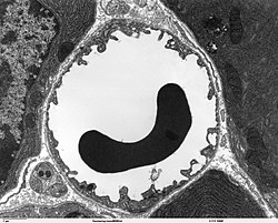
Capillaries
Capillaries (/ˈkæpɪlɛriz/ in US; /kəˈpɪləriz/ in UK) are the smallest of a body's blood vessels (and lymph vessels) that make up the microcirculation. Their endothelial linings are only one cell layer thick. These microvessels, measuring around 5 to 10 micrometres (µm) in diameter, connect arterioles and venules, and they help to enable the exchange of water, oxygen, carbon dioxide, and many other nutrients and waste substances between the blood and the tissues[3] surrounding them. Lymph capillaries connect with larger lymph vessels to drain lymph collected in the microcirculation.
During early embryonic development[4] new capillaries are formed through vasculogenesis, the process of blood vessel formation that occurs through a de novo production of endothelial cells which then form vascular tubes.[5] The term angiogenesis denotes the formation of new capillaries from pre-existing blood vessels and already present endothelial which divides.[6]
Blood
Red blood cells
The cytoplasm of erythrocytes is rich in hemoglobin, an iron-containing biomolecule that can bind oxygen and is responsible for the red color of the cells. The cell membrane is composed of proteins and lipids, and this structure provides properties essential for physiological cell function such as deformability and stability while traversing the circulatory system and specifically the capillary network.
In humans, mature red blood cells are flexible and oval biconcave disks. They lack a cell nucleus and most organelles, in order to accommodate maximum space for hemoglobin; they can be viewed as sacks of hemoglobin, with a plasma membrane as the sack. Approximately 2.4 million new erythrocytes are produced per second in human adults.[2] The cells develop in the bone marrow and circulate for about 100–120 days in the body before their components are recycled by macrophages. Each circulation takes about 60 seconds (one minute).[3] Approximately a quarter of the cells in the human body are red blood cells.[4][5] Nearly half of the blood's volume (40% to 45%) is red blood cells.
Red blood cells are also known as RBCs, red cells,[6] red blood corpuscles, haematids, erythroid cells or erythrocytes (from Greek erythros for "red" and kytos for "hollow vessel", with -cyte translated as "cell" in modern usage). Packed red blood cells (pRBC) are red blood cells that have been donated, processed, and stored in a blood bank for blood transfusion.
White blood cells
Platelets Platelets are tiny blood cells that help your body form clots to stop bleeding. If one of your blood vessels gets damaged, it sends out signals that are picked up by platelets. ... They also send out chemical signals to attract more platelets to pile onto the clot in a process called aggregation.

All white blood cells have nuclei, which distinguishes them from the other blood cells, the a nucleated red blood cells (RBCs) and platelets. Types of white blood cells can be classified in standard ways. Two pairs of broadest categories classify them either by structure (granulates or granulates) or by cell division lineage (myelitis cells or lymphoid cells). These broadest categories can be further divided into the five main types: necrophiliacs, eosinophils, basophils, lymphocytes, and monocles.[2] These types are distinguished by their physical and functional characteristics. Monocles and necrophiliacs are phagocyte. Further subtypes can be classified; for example, among lymphocytes, there are B cells, T cells, and NK cells.
The number of leukocytes in the blood is often an indicator of disease, and thus the WBC count is an important subset of the complete blood count. The normal white cell count is usually between 4 × 10⁹/L and 11 × 109/L. In the US this is usually expressed as 4,000 to 11,000 white blood cells per microliter of blood.[3] They make up approximately 1% of the total blood volume in a healthy adult,[4]making them substantially less numerous than the RBCs at 40% to 45%. However, this 1% of the blood makes a large difference to health, because immunity depends on it. An increase in the number of leukocytes over the upper limits is called leukocytes. It is normal when it is part of healthy immune responses, which happen frequently. It is occasionally abnormal, when it is neoplastic or autoimmune in origin. A decrease below the lower limit is called leukemia. It weakens the immune system.
Plasma Plasma is a state of matter. The three other common states of matter are solids, liquids and gases, so plasma is sometimes called the fourth state of matter. Plasma is created by adding energy to a gas so that some of its electrons leave its atoms. Platelets Platelets are tiny blood cells that help your body form clots to stop bleeding. If one of your blood vessels gets damaged, it sends out signals that are picked up by platelets. ... They also send out chemical signals to attract more platelets to pile onto the clot in a process called aggregation.

Subscribe to:
Posts (Atom)